Where Are the Main Arteries in the Body
7.4: Bloodline Vessels
- Page ID
- 30661

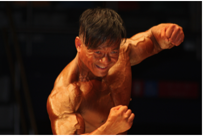
Why do bodybuilders have such prominent veins? Bulging muscles push coat veins closer to the skin. Couple that with a virtual lack of subcutaneous fat, and you have bulging veins as well as bulging muscles. Veins are one of three major types of line vessels in the circulatory system.
Types of Blood Vessels
Blood vessels are part of the cardiovascular arrangement that transports blood throughout the chassis. There are three major types of blood vessels: veins, arteries, and capillaries.

Arteries are defined as blood vessels that carry blood away from the heart. Blood flows through and through arteries for the most part because it is under pressure from the pumping action of the heart. IT should represent noted that coronary arteries, which supply heart muscle cells with pedigree, jaunt toward the heart but not as region of the blood feed that travels through the chambers of the heart. Most arteries, including coronary arteries, carry oxygenated blood, but there are a few exceptions, most notably the pulmonic arteria. This artery carries deoxygenated blood from the affection to the lungs, where it picks up oxygen and releases carbon dioxide. In virtually all other arteries, the haemoglobin in red blood cells is extremely saturated with oxygen (95-100 percent). These arteries distribute oxygenated blood to tissues throughout the body.
The largest arterial blood vessel in the body is the aorta, which is connected to the heart and extends down into the abdomen (Figure \(\PageIndex{2}\)). The aorta has aggressive, oxygenated blood pumped up directly into it from the unexhausted ventricle of the heart. The aorta has many branches, and the branches subdivide repeatedly, with the subdivisions growth smaller and smaller in diam. The smallest arteries are called arterioles.
Veins
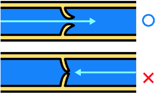
Veins are characterised Eastern Samoa blood vessels that carry blood toward the heart. Roue traveling through veins is non under pressure from the beating heart. It gets assist moving along by the squeezing action of emaciated muscles, for instance, when you walk or breathe. It is also prevented from flowing backward by valves in the larger veins, as illustrated in Material body \(\PageIndex{3}\). Veins are called capacitance blood vessels because the majority (about 60 percent) of the body's entire volume of pedigree is contained within veins.
Most veins post deoxygenated blood, but thither are a few exceptions, including the four pulmonary veins. These veins carry oxygenated blood from the lungs to the meat, which then pumps the blood to the rest of the organic structure. In near all other veins, hemoglobin is relatively unsaturated with oxygen (about 75 per centum).
The two largest veins in the body are the precava, which carries blood from the upper body directly to the proper atrium cordis, and the cheapjack vena cava, which carries stoc from the lower body now to the rightist atrium. The tinny vena cava is labeled in the enter below. The superior vein cava is not labeled in Anatomy \(\PageIndex{4}\) but is clearly visible entering the right atrium cordis. Ilk arteries, veins form a complex, forking organisation of larger and smaller vessels. The smallest veins are called venules. They receive ancestry from capillaries and enthrall it to larger veins. Each venule receives blood from multiple capillaries.

Capillaries
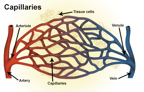
Capillaries are the smallest blood vessels in the cardiovascular system of rules. They are indeed pocket-sized that only one RBC at a time can mash through and through a capillary tubing, and so only the bloody blood mobile phone deforms. Capillaries colligate arterioles and venules, as shown in Picture \(\PageIndex{5}\). Capillaries generally form a ramate network of vessels, called a capillary bed, that provides a turgid area for the commute of substances between the blood and surrounding tissues.
Structure of Blood Vessels
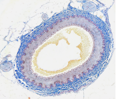
All blood vessels are basically hollow tubes with an internal quad, called a lm, through which blood flows. The lumen of an artery is shown in crosswise in the photomicrograph below. The breadth of blood vessels varies, merely they totally have a lumen. The walls of blood vessels differ depending on the case of vessel. In widespread, arteries and veins are more than similar to indefinite another than capillaries in the structure of their walls.
Walls of Arteries and Veins
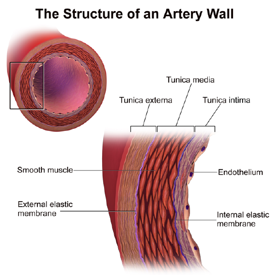
The walls of both arteries and veins have three layers: the tunica intima, tunica media, and tunica adventitia. You keister get word the three layers for an arterial blood vessel in Figure \(\PageIndex{7}\).
- The tunica intima is the internal layer of arteries and veins. It is also the thinnest layer, consisting of a single layer of endothelial cells encircled by a thin level of connective tissues. It reduces friction between the blood and the inside of the blood vessel walls.
- The tunica media is the halfway layer of arteries and veins. In arteries, this is the thickest layer. It consists mainly of viscoelastic fibers and connective tissues. In arteries, this is the thickest layer because it also contains involuntary muscle tissues, which ensure the diam of the vessels.
- The adventitia externa (also called tunica adventitia) is the outmost stratum of arteries and veins. It consists of connective weave and as wel contains nerves. In veins, this is the thickest level. In general-purpose, the tunica externa protects and strengthens vessels and attaches them to close structures.
Capillary Walls
The walls of capillaries consist of little more than a one-member layer of epithelial cells. Beingness just unrivaled electric cell thick, the walls are well suited for the exchange of substances between the blood inside them and the cells of surrounding tissues. Substances including water, oxygen, glucose, and opposite nutrients as good as waste products such as CO2 can pass quickly and easily through and through the extremely thin walls of capillaries.
Blood Insistency
The blood in arteries is normally under pressure because of the rhythmical of the heart. The pressure is highest when the heart and soul contracts and pumps out roue, and last when the heart relaxes and refills with rakehell. (You can feel this variation in squeeze in your wrist or cervix when you look your pulse.) Line of descent pressure is a criterion of the force that blood exerts on the walls of arteries. IT is generally measured in millimeters of mercury (millimetre Hg) and expressed as a double routine: a high number for systolic pressure when the ventricles contract; and a lower number for diastolic pressure sensation when the ventricles relax. Normal blood pressure is in the main defined equally to a lesser extent than 120 mm Hg (systolic)/80 millimeter of mercury (diastolic) when measured in the arm at the level of the heart. It decreases American Samoa blood flows farther away from the essence and into smaller arteries.
Every bit arteries grow smaller, in that location is increasing resistivity to blood flow finished them because of the detrition of the line against the arterial walls. This resistance restricts blood flow rate so less blood reaches smaller, downstream vessels, thus reducing profligate coerce before the blood flows into the tiniest vessels, the capillaries. Without this reduction in blood pressure, capillaries would not be able to hold up the pressure level of the blood without bursting. Away the prison term blood flows through the veins, it is below very little pressure. The pressure of blood against the walls of veins is always about the cookie-cutter and ordinarily no to a higher degree 10 mm Hg.
Vasoconstriction and Vasodilation
Smooth muscles in the walls of arteries bathroom contract or relax to cause vasoconstriction (constricting of the lm of blood vessels) or vasodilation (widening of the lumen of blood vessels). This allows the arteries — especially the arterioles — to contract or relax as needed to help regulate blood pressure. In this heed, the arterioles play same an changeable nozzle on a garden hose. When they dogmatical, the increased friction with the arterial walls causes less blood to menstruation downriver from the narrowing, ensuant in a drop in descent imperativeness. These actions are controlled by the involuntary nervous system in response to pressure-responsive sensorial receptors in the walls of bigger arteries.
Arteries can also dilate or constrict to help regulate body temperature by allowing more Oregon less blood to flow from the ardent physical structure nucleus to the body's opencut. In addition, vasoconstriction and vasodilation play roles in the fight-operating theatre-flight response, under the check of the sympathetic excited system. For example, vasodilation allows more stoc to flow to wasted muscles and vasoconstriction reduces blood flow to digestive variety meat.
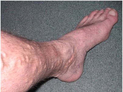
The chunky appearing of this man's leg is caused past varicose veins. Do you have varicose veins? If you do, you may admiration whether they are a sign of a significant health problem. You whitethorn besides wonder whether you should have them treated, and if indeed, what treatments are purchasable. As is usually the case, when information technology comes to your health, "knowledge is office."
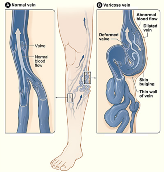
First, the "back story:" varicose veins are veins that have become enlarged and distorted because their valves have become ineffective (see Anatomy \(\PageIndex{8}\)). As a consequence, blood pools in the veins and stretches them out. Unhealthy veins occur most frequently in the superficial veins of the legs, but they may also occur in other parts of the body. They are most common in old adults, females, and the great unwashe WHO induce a family history of the condition. Fleshiness and pregnancy also increase the risk of developing varicose veins. A job that requires standing for long periods of time, chronic irregularity, and long-term alcohol uptake are additional risk factors.
Varicose veins usually are not serious. In many people, they are only a cosmetic issue. However, in severe cases, unhealthy veins may cause ail and other problems. E.g., the mannered leg(s) may palpate heavy and achy, especially after long periods of standing. Ankles Crataegus laevigata become swollen away the end of the daytime. Minor injuries may leech to a higher degree modal. The skin over varicosity Crataegus oxycantha become red, bone-dry, and itchy. In identical severe cases, skin ulcers English hawthorn develop.
If you are related to about varicose veins, call them to the attention of your doctor, World Health Organization terminate determine the best course of accomplish for your case. There are many potential treatments for varicose veins. About of the treatments have potential adverse side effects; and with many of the treatments, varicose veins may return. Which discourse is best for a conferred patient depends in part on the severity of the condition.
- If varicose veins are non serious, then conservative discourse options may be recommended. These include avoiding lasting or sitting for long periods, frequently elevating the legs, and tiring proportional compression stockings.
- For more serious cases, less fusty but non-surgical options may be advised. These include sclerotherapy, in which medicine is injected into the veins to make them shrink. Another non-postoperative approach is endovenous thermal excision. In this character of treatment, laser light, radio-frequency energy, or steam is used to heat the walls of the veins, causing them to shrink and collapse.
- For the well-nig serious cases, surgery may be the good option. The near invasive surgery is vein stripping, in which all surgery part of the main bole of a vein is knotted soured and removed from the pegleg spell the patient is low general anesthesia. In a less invasive surgery, named ambulatory phlebectomy, short segments of a vein are remote through tiny incisions under local anesthesia.
Review
- What are the rakehell vessels? Key the terzetto better types of blood vessels.
- Describe arteries. Identify the largest artery in the body.
- How are veins characterised? What are the two largest veins in the body?
- Compare and contrast how descent moves through arteries and veins.
- What are capillaries, and what is their function?
- Compare and contrast the structure of the walls of arteries, veins, and capillaries.
- What is descent coerce, and how is it expressed? What blood pressure is considered sane?
- Identify the functions of vasoconstriction and vasodilation of arteries.
- Does the blood in virtually veins have whatever oxygen at all? Explain your result.
- True or False. Only unity RBC can pass off through the lumen of a hairlike at a acknowledged clip.
- True or False. The arteria pulmonalis carries oxygenated stoc.
- Which tissue in blood vessels is responsible vasodilation and vasoconstriction? Where is it located?
- The blood pressure level at the arterioles is generally _________ the blood pressure at the aorta.
A. lower than
B. higher than
C. the same as
D. non related
- Explicate why it is key that the walls of capillaries are very thin.
- Most of the blood in the body is in the:
A. Capillaries
B. Arteries
C. Heart
D. Veins
Search More
Where Are the Main Arteries in the Body
Source: https://bio.libretexts.org/Courses/Community_College_of_Vermont/Human_Biology_%28Gabor_Gyurkovics%29/07:_Cardiovascular_System/7.04:_Blood_Vessels
0 Response to "Where Are the Main Arteries in the Body"
Publicar un comentario